A New Super-Resolution Microscopy Technique Lets Scientists Study Cells In Real Time
Our take
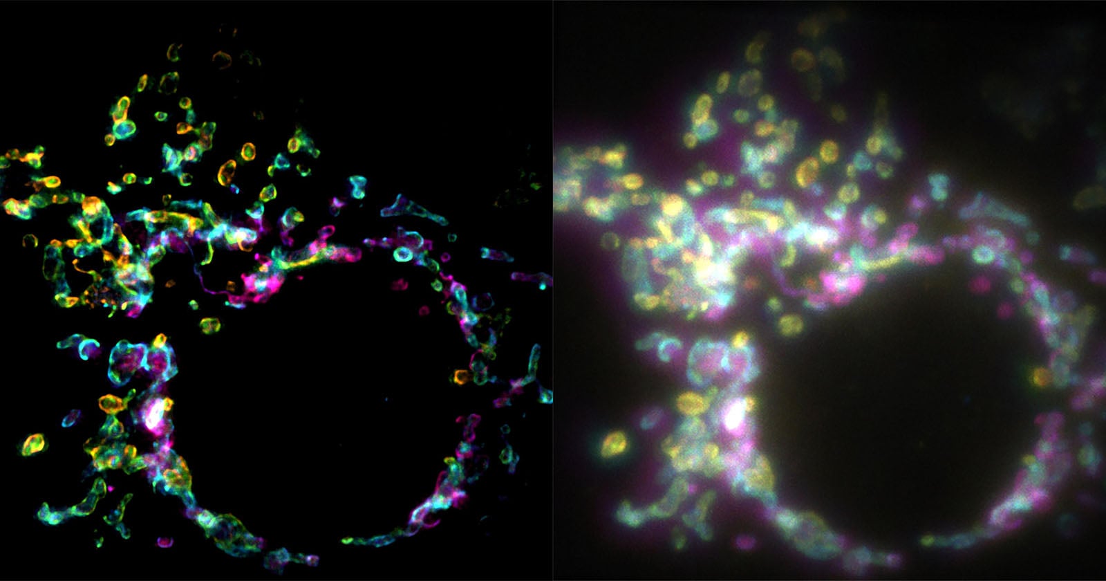
The unveiling of a new super-resolution microscopy technique capable of generating high-resolution images from a single exposure marks a significant leap forward in cellular biology, and one that resonates deeply with our audience’s appreciation for detail and innovation. For years, scientists have strived to overcome the diffraction limit of light, a fundamental barrier preventing the visualization of cellular structures with clarity. Existing super-resolution methods, while groundbreaking, often involve complex protocols, multiple exposures, and intricate data processing – a considerable hurdle for many labs. This new technique promises to simplify that process considerably, potentially democratizing access to this powerful imaging modality. It’s akin to the evolution from early photographic processes to the instant gratification of digital photography; a shift towards accessibility and immediacy. We've previously explored the power of capturing detail in other mediums, like the glowing aesthetic of Cinestill film A Glowing Endorsement of CineStill’s Super Popular 800T Film and how a single image can achieve viral fame What Happens When Your Photograph Becomes a Viral Meme?, and this advancement speaks to the same underlying desire to see and understand the world with greater fidelity.
The ability to observe cellular processes in real-time, with such remarkable clarity, unlocks a wealth of possibilities. Researchers can now potentially track dynamic events within cells – protein interactions, organelle movements, and signaling cascades – with unprecedented precision. This could accelerate discoveries in areas ranging from cancer biology and neurodegenerative diseases to drug development and regenerative medicine. Consider the implications for personalized medicine; the ability to analyze cellular behavior with this level of detail could lead to more targeted therapies and improved diagnostics. The technique’s simplicity also suggests it could be adapted for use in educational settings, providing students with a more immersive and engaging learning experience. This isn't just about seeing *more*; it's about seeing *differently* and gaining insights previously inaccessible. The potential for visual storytelling within scientific research is amplified as well; imagine the artistic potential of capturing these intricate cellular landscapes in stunning detail.
Beyond the immediate scientific impact, this advancement highlights the ongoing convergence of disciplines – physics, engineering, and biology – driving innovation at the nanoscale. The development of this technique undoubtedly relied on breakthroughs in optics, materials science, and computational algorithms. It’s a testament to the power of interdisciplinary collaboration and the relentless pursuit of knowledge. The field of microscopy itself is undergoing a renaissance, with new technologies constantly pushing the boundaries of what's visible. We've seen this evolution mirrored in other areas of technology, from the iterative improvements in camera technology, as discussed in our recent podcast Did the New PEN Impress You? Plus Apple Adds Aperture | The PetaPixel Podcast, to the continuous refinement of image processing techniques. This new super-resolution method fits seamlessly into that narrative of relentless progress.
Looking ahead, the crucial question becomes: how rapidly will this technique be adopted by the broader scientific community? Will it truly live up to its promise of simplifying super-resolution imaging and making it accessible to a wider range of researchers? The initial reports are promising, but the long-term impact will depend on factors such as cost, ease of implementation, and compatibility with existing experimental workflows. Regardless, this development represents a significant milestone in our ability to visualize the fundamental building blocks of life, opening a new window into the intricate world within our cells and inviting us to explore its beauty and complexity with a fresh perspective.
A new fluorescence microscopy technique promises to create super-resolution images from a single exposure and give scientists a remarkable look inside cells.
Read on the original site
Open the publisher's page for the full experience