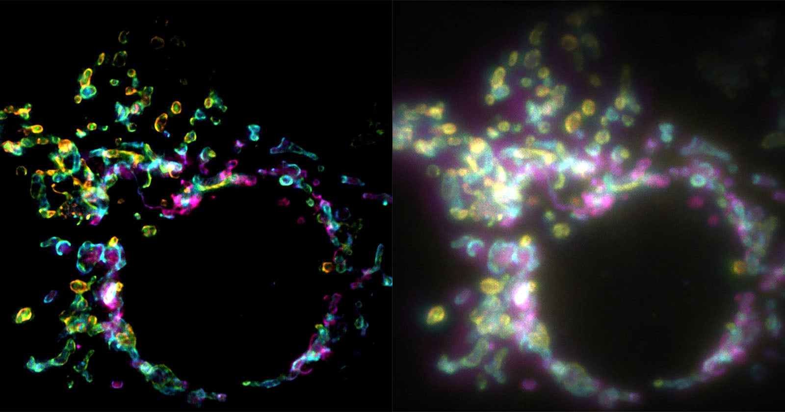
There is something quietly profound about a tool that lets us watch life while it is still happening. The new fluorescence microscopy technique promises super-resolution images from a single exposure, which means scientists can now study cells in real time without the usual trade-off between speed and clarity. For years, capturing the inner workings of a cell in crisp detail required freezing the moment in time, sacrificing the living, breathing context of the process. This approach changes that equation, and it is hard not to feel a spark of wonder at the implications.
We spend so much time at the intersection of art and technology here, whether we are reviewing a Refined and Accessible: The Nikon Z5 IIC—A Full-Frame Camera, Reimagined or marveling at the latest Vivo X500: A Curated Lens for the Art of Life. This feels like a cousin to that same obsession with capturing moments, except the subject is not a landscape or a portrait but the very machinery of life itself. What excites us is not just the sharper image, but the access it grants. A single exposure means less phototoxicity, less disturbance to the sample, and more opportunities to witness cellular events as they unfold. For researchers, this is not a small upgrade; it is a new way of seeing.
Our honest take is that this is less about the microscope itself and more about what it unlocks for the people using it. Biologists have long been forced to choose between resolution and live imaging, and this technique quietly dissolves that dilemma. For a reader who might ask us whether this matters beyond the lab, we would say this: every major medical breakthrough, from targeted therapies to regenerative medicine, begins with understanding how cells behave. Tools like this do not just make science more efficient; they make it more intuitive. There is an artistry to watching a cell divide in real time, to seeing the subtle choreography of proteins moving through a membrane, that a static image can never convey. It is immersive in the truest sense.
The practical takeaway here is simple: this is a tool that will likely shorten the distance between observation and understanding. We would tell a curious reader to watch how this technique is adopted in the coming years, not because the technology is flashy, but because it quietly removes a barrier that has slowed discovery for decades. The question is not whether scientists will use it, but what they will choose to look at first. In a world where we are constantly chasing sharper images, from our cameras to our microscopes, the real magic is always in what we choose to observe.